Hip Muscles Diagram / Muscle Synergies Of The Hip And Pelvis Rayner Smale / Press into the feet, lengthening the legs to press the hips up toward the ceiling.
Hip Muscles Diagram / Muscle Synergies Of The Hip And Pelvis Rayner Smale / Press into the feet, lengthening the legs to press the hips up toward the ceiling.. The different bursae of the hip region (trochanteric, ischial and. The muscular system is responsible for the movement of the human body. They can be divided into three main groups: Human anatomy for muscle, reproductive, and skeleton. The hip muscles cover the hip joint as a muscle sheath.
It therefore serves the artist as a dependable visual landmark for the location of muscular forms. Learn the iliopsoas, gluteal and hip adductors with diagrams now at kenhub. Muscle and tendon anatomy of the hip (adductors, gluteal muscles (or buttocks), hamstring muscles, femoral muscle quadrices). It joins the lower limb to the pelvic girdle. Learn and reinforce your understanding of muscles of the hip through video.
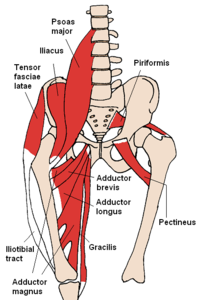
Muscles Of The Hip Wikipedia from upload.wikimedia.org Bursae of the lower limb: Diagram representing the anterior view of the muscle groups adductor brevis, adductor longus and adductor magnus. The hip and pelvic muscles include: The following diagram illustrates the actions of the terms adduction, abduction, flexion and extension at the different joints. If you are starting to feel hip pain or stiffness, you'll want to know hip anatomy: Knee assessment and hip mechanics learn how hip and pelvis. It therefore serves the artist as a dependable visual landmark for the location of muscular forms. The different bursae of the hip region (trochanteric, ischial and.
Related online courses on physioplus.
Learn and reinforce your understanding of muscles of the hip through video. The external rotation of the hip helps people get into cars, pitch baseballs, and do a variety of other activities. Other muscles also assist in the abduction of the thigh at the hip joint, but they do not belong to the abductor group. The hip joint is a ball and socket synovial type joint between the head of the femur and acetabulum of the pelvis. Diagram representing the anterior view of the muscle groups adductor brevis, adductor longus and adductor magnus. See more ideas about muscle diagram, medical anatomy, muscle anatomy. Most modern anatomists define 17 of these muscles, although some additional muscles may sometimes be considered. Bones in human anatomy, the hip flexors are a group of skeletal muscles that act to flex the femur (thigh bone) hip muscles diagram. The sacrum bone is almost always noticeable, no matter what the body type, because it is not covered with muscles or substantial fatty tissue. Their main function is contractibility. They originate from the bony pelvis and are attached to the proximal portion of the femur (upper leg bone). The hip muscles cover the hip joint as a muscle sheath. Learn vocabulary, terms and more with flashcards, games and other study tools.
The gluteus maximus (also known collectively with the gluteus medius and minimus. Press into the feet, lengthening the legs to press the hips up toward the ceiling. Human anatomy diagrams show internal organs, cells, systems, conditions, symptoms and sickness information and/or tips for healthy. See more ideas about muscle diagram, medical anatomy, muscle anatomy. If you are starting to feel hip pain or stiffness, you'll want to know hip anatomy:
3 Superficial And Deep Posterior Hip Muscles Diagram Quizlet from o.quizlet.com 25.09.2020 · the hip muscles encompass many muscles of the hip and thigh whose main function is to act on the thigh at the hip joint and stabilize the pelvis.without them, walking would be impossible. These two muscles produce lateral rotation at the hip and are innervated by the obturator internus and quadratus femoris nerves. Muscles, connected to bones or internal organs and blood vessels, are in charge for movement. Attached to the bones of the skeletal system are about 700 named. This diagram depicts muscles in hip area 744×1208. Comprehensive information about hip joint anatomy including muscles, tendons, ligaments, bones, bursae, skeletal structure and joint capsules. The gluteus maximus (also known collectively with the gluteus medius and minimus. This is the largest of the three compartments of the thigh.
There are 21 different muscles that cross the hip joint.
A hip flexor and mild hip lateral rotator. These two muscles produce lateral rotation at the hip and are innervated by the obturator internus and quadratus femoris nerves. The anatomy of the fascia lata and iliotibial tract. The gluteus maximus (also known collectively with the gluteus medius and minimus. The movements that can be carried out at the hip joint are listed below, along with the principle muscles responsible for each action Human anatomy for muscle, reproductive, and skeleton. Want to learn more about it? Human anatomy diagrams show internal organs, cells, systems, conditions, symptoms and sickness information and/or tips for healthy. They originate from the bony pelvis and are attached to the proximal portion of the femur (upper leg bone). Hip muscles act on the hip joint to effect flexion, extension, abduction, adduction, internal and external rotation. They can be divided into three main groups: Muscle and tendon anatomy of the hip (adductors, gluteal muscles (or buttocks), hamstring muscles, femoral muscle quadrices). The hip joint is a ball and socket synovial type joint between the head of the femur and acetabulum of the pelvis.
If you are starting to feel hip pain or stiffness, you'll want to know hip anatomy: The muscular system is responsible for the movement of the human body. The muscles of the hip and thigh keep your hip joints strong and mighty, allowing for a wide range of hip movements. Most modern anatomists define 17 of these muscles, although some additional muscles may sometimes be considered. Want to learn more about it?
Understand Hip Anatomy Muscles For Yoga Jason Crandell Yoga from www.jasonyoga.com Hip muscles diagram, learn more about hip muscles diagram. Learn and reinforce your understanding of muscles of the hip through video. Feel the spine being pulled in opposite directions as you press the head. The anatomy of the fascia lata and iliotibial tract. It therefore serves the artist as a dependable visual landmark for the location of muscular forms. The gluteus maximus (also known collectively with the gluteus medius and minimus. Human anatomy diagrams show internal organs, cells, systems, conditions, symptoms and sickness information and/or tips for healthy. Knee assessment and hip mechanics online course:
Learn and reinforce your understanding of muscles of the hip through video.
Muscle and tendon anatomy of the hip (adductors, gluteal muscles (or buttocks), hamstring muscles, femoral muscle quadrices). The movements that can be carried out at the hip joint are listed below, along with the principle muscles responsible for each action In this article we describe the hip and thigh muscles. The anatomy of the fascia lata and iliotibial tract. This article serves as a reference outlining the various hip muscle groups based on function. In human anatomy, the muscles of the hip joint are those muscles that cause movement in the hip. The hip and pelvic muscles include: The muscular system is responsible for the movement of the human body. Diagram representing the anterior view of the muscle groups adductor brevis, adductor longus and adductor magnus. There are 21 different muscles that cross the hip joint. Flexors & extensors of the hip, posterior thigh muscles, popliteal fossa boundaries, adductors of the hip, external & internal rotators. Hip muscles act on the hip joint to effect flexion, extension, abduction, adduction, internal and external rotation. Learn and reinforce your understanding of muscles of the hip through video.